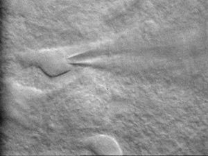
The MOBs team was created by Karim Benchenane (CR1 CNRS) as a part of the Brain Plasticity Unit at the ESPCI since 2012. The head of the team has an expertise in the role of sleep in memory consolidation based on electrophysiology in freely moving rodents, and Thierry Gallopin (MCF ESPCI) is an expert of patch clamp study who has a strong expertise in the activity of brain structures responsible for sleep/wake regulation and in the physiology of cortical interneurons.
The aim of the MOBs team is to study the brain states related to the different brain oscillatory modes and their role in memory processing, based on in vivo and ex-vivo approaches.
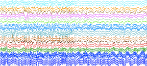
The brain is not a passive structure that receives, processes, encodes and stores information. On the contrary, it shows self-generated oscillations at different frequencies during periods that have been defined as brain states. The different brain states are related to different vigilance states (wakefulness, REM sleep, SWS) and each states is considered to be related to different steps in memory processing (encoding, consolidation). We intent to study the brain states, their related brain oscillatory modes and their role in memory processing. This will be done in a main project that is funded by an ERC consolidator Grant and in three additional smaller projects and will be achieved by using electrophysiology in freely moving mice in vivo and in slice ex-vivo, both approaches being coupled with optogenetic tools.
Past achievements

It is now commonly accepted that sleep plays a crucial role in the memory consolidation process. Numerous studies have shown that some of the positive effect of sleep on consolidation relies on the reactivations of brain activity from previous experiences, occurring during slow-waves sleep (SWS).
During behavior, hippocampal place cells discharge when the animal is in particular locations of the environment (called place fields). The activation sequences of hippocampal place cells corresponding to the trajectories previously followed are ‘replayed’ during SWS. Yet, no study ever addressed a causal role of replay on behavior or memory.
To address this issue, we developed a brain- computer interface to detect in real-time the action potentials of a place cell and automatically trigger (after each detection) stimulations of the medial forebrain bundle, known to simulate reward in rodents and create a memory that the place field was associated with a pleasurable experience. This stimulation during sleep led to the mice going directly to the place field of the neuron when animals they woke up, thus demonstrating that the association between place and reward had been created during sleep (de Lavilléon et al. Nat Neurosci 2015).
This experiment provides direct evidence that place cell activity indeed conveys spatial information, even during sleep, further supporting the concept of reactivation. This work made a huge step beyond correlative studies and showed a causal role of place cells in navigation and was highly cited in the popular and scientific press.

Since several lines of evidence show that astrocytes can regulate cortical slow oscillations during sleep, we investigated the role of connexins (Cx), the main constituent of gap junctions and hemichannels) in sleep physiology.
We first analyzed sleep-like oscillation of the membrane potential of mitral cells in olfactory bulb ex-vivo in slices from wild-type and double knock-out mice (dKO) for astrocytic Cx43 and Cx30. Interestingly, the amplitude of these oscillations were decreased in slices from dKO mice (Roux et al., J Neurosci 2015).
We also analyzed sleep physiology in vivo by multi-site recordings (in several brain structures including the olfactory bulb) in dKO mice, supporting a role of astrocytes in sleep.
Thanks to the recordings performed in the olfactory bulb we showed that the gamma (40-80 Hz) oscillation in the olfactory bulb is strongly decreased during sleep (both during non-REM and REM sleep) compared to the awake state. This decrease was strong enough so that we could use it to perform sleep scoring with 92% accuracy. This shows that we can define sleep by using only brain signals, while previous methods rely on indirect and difficult measures of wakefulness such as motor activity (video tracking or EMG).
Finally, we decided to characterize in greater detail the structure of non-REM sleep. While non-REM sleep is traditional subdivided in humans into 3 stages called N1, N2 and N3, in mice non-REM sleep is often considered as a homogenous state. We showed that this is not the case and we were able to differentiate 3 states that share the same characteristics with he human non-REM sleep substages. Importantly the evolution of the newly defined sleep states over sleep cycles is similar in rodent and human, as well as the probability of transition between these stages. These results will improve the transferability of the results obtained in mice to human by using comparable methodologies of sleep scoring.

The research activities headed by Thierry Gallopin were focused on the understanding of the cellular and molecular mechanisms responsible for the initiation and maintenance of non-REM sleep in mice.
One of the major finding of Gallopin’s team was the identification that glucose may participate in the mechanisms of sleep promotion and/or consolidation. Indeed, by combining in vitro and in vivo experiments, they demonstrated that glucose likely contributes to sleep onset facilitation by selectively increasing the excitability of sleep-promoting neurons in VLPO by direct and indirect pathways.
The first one requires the intra-neuronal metabolization of glucose to ATP, which in turn inhibits ATP-sensitive potassium (KATP) channels leading to a depolarization of the cell (Varin et al., 2015).
The second mechanism implies glucose uptake by astrocytes which lead to extracellular adenosine release that depolarize neurons through A2AR activation (Scharbarg et al., 2016). This glucose-induced excitation of sleep-promoting neurons should therefore be involved in the drowsiness that one feels after a high-sugar meal. This novel mechanism regulating the activity of VLPO neurons reinforces the fundamental and intimate link between sleep and metabolism.
Given the critical role of the VLPO in sleep induction and maintenance, the characterization of its neuronal populations is essential for a better understanding of their role in sleep regulation. Based on their strong background in the multiparametric characterization of the GABAergic neuronal diversity in the neocortex (Perrenoud et al., 2012a; 2012b; 2013) they defined the existence of several distinct cell subpopulations in the VLPO (Dubourget et al., 2016; Sangare et al., 2016). These different types of cells suggest a complex neuronal network organization within the VLPO with specific functions on sleep regulation.
Finally, in order to better understand the mechanisms of sleep pressure and the consequences of the lack of sleep, we have developed different techniques of sleep deprivation. These approaches allowed them to define the impact of deprivation and / or sleep restriction at the cellular, behavioral and cognitive levels (Chennaoui et al., 2015).


